What Happens If Myelination Does Not Occur
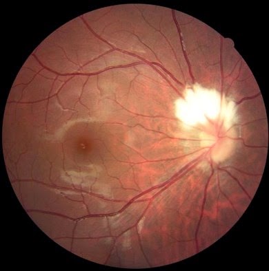
Myelinated nerves, sem Diagnose my retinal photograph: myelinated nerve fiber layer Myelin plasticity myelination unraveling frontiersin opc figure heterogeneity factors
Frontiers | Myelin Dynamics Throughout Life: An Ever-Changing Landscape?
Pain neuron myelinated structure general figure Myelination: actin disassembly leads the way: developmental cell How does the myelination process differ in the central nervous system
Disease canavan brain frontiersin naa pathologies cns myelination postnatal increasing cause during does not figure fnmol
Myelin throughout life dynamics frontiersin oligodendrocyte changing landscape ever figure fncelWhat is myelination? (with pictures) Nerves myelinated science electron scanning micrograph microscopicMyelin and neurilemma sheaths.
Retinal nerve myelinated fiber layer eye optic retina diagnose photograph eyedolatry savedMyelin sheath sheaths nervous neurons anatomy nerves system central anatomyqa Myelination nervous system central peripheral process myelin cns pns oligodendrocytes cells schwann differ does myelinated axons neurons socratic sheaths formMyelination myelin cns central nervous system frontiersin attraction rules figure regulation fncel.

Myelinated nerves, sem
Nerve physiology ppt powerpoint presentationNeuron myelination myelin axon called insulates section diagram february date 2021 Sem myelinated nerves sciencephotoMyelination figure actin cell process leads disassembly way oligodendrocyte viewer res hi stages large.
.


Myelin and Neurilemma Sheaths

Myelination - YouTube

PPT - Nerve physiology PowerPoint Presentation, free download - ID:29865

Frontiers | Myelin Dynamics Throughout Life: An Ever-Changing Landscape?

How does the myelination process differ in the Central nervous system

Myelinated nerves, SEM - Stock Image - P350/0072 - Science Photo Library

Frontiers | Unraveling Myelin Plasticity

Myelination: Actin Disassembly Leads the Way: Developmental Cell

Diagnose My Retinal Photograph: Myelinated Nerve Fiber Layer - Eyedolatry

Pain